
3D foot scanning for custom orthotics
A clinical foot scanner
in your pocket.
Foot Camera turns an iPhone or iPad with Face ID into a 3D foot scanner. Capture a foot in under a minute, watch the coverage build live, and send lab-ready files without leaving the treatment room.
Requires an iPhone or iPad with a Face ID camera, running iOS 18 or later.
~100k
measured surface points per scan
30–45 s
to capture one foot
1–2 mm
depth accuracy at working range
Built for the treatment room, not the lab bench
Everything the scan needs happens on the device, in front of the patient — capture, guidance, quality checks and review.
TrueDepth capture, no rig
The same sensor that powers Face ID projects roughly 30,000 infrared dots onto the foot and measures the distance to every point on the surface, thirty times a second — accurate to about 1–2 mm at working range. No frame, no calibration target, no consumables.
Watch the scan paint on
Coverage builds on screen as you move around the foot: yellow where the surface is newly seen, green once it is well observed. Colour and sound cues hold you in the 28–38 cm sweet spot, so you know the scan is good before you stop.
Quality checked, frame by frame
Every frame is tested against motion and alignment gates as it is captured. If the app ever loses confident lock on the foot it stops cleanly and offers a retake — it will never quietly hand the lab a distorted model.
Lab-ready and offline-first
Finished scans export in your lab’s standard formats, in metres, with true colour — so tender-area circles and bisection lines drawn on the foot are captured too. Uploads queue on the device and go out when there is signal.
How it works
Three steps, one appointment. Nothing to post, cast or courier.
- 1
Create the order
Patient details and which feet are being scanned. Practitioners sign in with a one-time code sent to their work email — no passwords to manage.
- 2
Scan each foot
Hold the phone about a hand’s length from the foot and move slowly around the sole, sides and heel. A foot takes 30–45 seconds.
- 3
Review, then send
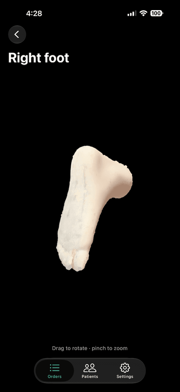
Inspect the 3D model on screen — rotate and zoom — before accepting it. Accepted scans upload automatically and reach the lab in its own format.
A scan you can defend
The reconstruction engine tracks the camera against the model it has already built, rejecting any frame that implies an impossible movement. Rather than blending a bad alignment into the model, it stops. Every scan also carries a diagnostic record — device, software version, tracking quality and the settings in force — so a question about a particular scan can be answered after the fact.
- Scans, patient details and files are held on servers in the European Union.
- Practitioners sign in with a one-time code; there are no shared passwords.
- Clinic staff see their own clinic’s patients; labs see only the orders sent to them.
- Poor connectivity is expected — scans queue on the device and upload later.
In limited release with partner clinics
Accounts are issued to clinics and laboratories directly. Get in touch to talk about your practice.